Search

Categories

    • categories-img Jacket, Women
    • categories-img Woolend Jacket
    • categories-img Western denim
    • categories-img Mini Dresss
    • categories-img Jacket, Women
    • categories-img Woolend Jacket
    • categories-img Western denim
    • categories-img Mini Dresss
    • categories-img Jacket, Women
    • categories-img Woolend Jacket
    • categories-img Western denim
    • categories-img Mini Dresss
    • categories-img Jacket, Women
    • categories-img Woolend Jacket
    • categories-img Western denim
    • categories-img Mini Dresss
    • categories-img Jacket, Women
    • categories-img Woolend Jacket
    • categories-img Western denim
    • categories-img Mini Dresss

Filter By Price

$
-
$

Dietary Needs

Top Rated Product

product-img product-img

Modern Chair

$165.00
product-img product-img

Plastic Chair

$165.00
product-img product-img

Design Rooms

$165.00

Brands

  • Wooden
  • Chair
  • Modern
  • Fabric
  • Shoulder
  • Winter
  • Accessories
  • Dress

Welcome and thank you for visiting us. For any query call us on 0799 626 359 or Email [email protected]

Offcanvas Menu Open

Shopping Cart

Africa largest book store
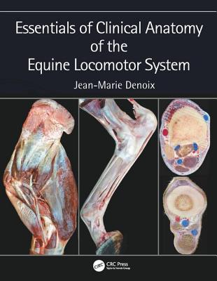
Sub Total:

Search for any Title

Essentials of Clinical Anatomy of the Equine Locomotor System

By: Jean-Marie Denoix (Author) , Jean-Marie Denoix (Author) , Jean-Marie Denoix (Author) , Jean-Marie Denoix (Author) , Jean-Marie Denoix (Author) , Jean-Marie Denoix (Author) , Jean-Marie Denoix (Author) , Jean-Marie Denoix (Author) , Jean-Marie Denoix (Author) , Jean-Marie Denoix (Author) , Jean-Marie Denoix (Author) , Jean-Marie Denoix (Author) , Jean-Marie Denoix (Author) , Jean-Marie Denoix (Author) , Jean-Marie Denoix (Author) , Jean-Marie Denoix (Author) , Jean-Marie Denoix (Author) , Jean-Marie Denoix (Author) , Jean-Marie Denoix (Author) , Jean-Marie Denoix (Author) , Jean-Marie Denoix (Author) , Jean-Marie Denoix (Author) , Jean-Marie Denoix (Author) , Jean-Marie Denoix (Author) , Jean-Marie Denoix (Author) , Jean-Marie Denoix (Author) , Jean-Marie Denoix (Author) , Jean-Marie Denoix (Author) , Jean-Marie Denoix (Author) , Jean-Marie Denoix (Author) , Jean-Marie Denoix (Author) , Jean-Marie Denoix (Author) , Jean-Marie Denoix (Author) , Jean-Marie Denoix (Author) , Jean-Marie Denoix (Author) , Jean-Marie Denoix (Author) , Jean-Marie Denoix (Author) , Jean-Marie Denoix (Author) , Jean-Marie Denoix (Author) , Jean-Marie Denoix (Author) , Jean-Marie Denoix (Author) , Jean-Marie Denoix (Author) , Jean-Marie Denoix (Author) , Jean-Marie Denoix (Author) , Jean-Marie Denoix (Author) , Jean-Marie Denoix (Author) , Jean-Marie Denoix (Author) , Jean-Marie Denoix (Author) , Jean-Marie Denoix (Author) , Jean-Marie Denoix (Author) , Jean-Marie Denoix (Author) , Jean-Marie Denoix (Author) , Jean-Marie Denoix (Author) , Jean-Marie Denoix (Author) , Jean-Marie Denoix (Author) , Jean-Marie Denoix (Author) , Jean-Marie Denoix (Author) , Jean-Marie Denoix (Author) , Jean-Marie Denoix (Author) , Jean-Marie Denoix (Author) , Jean-Marie Denoix (Author) , Jean-Marie Denoix (Author) , Jean-Marie Denoix (Author) , Jean-Marie Denoix (Author) , Jean-Marie Denoix (Author) , Jean-Marie Denoix (Author) , Jean-Marie Denoix (Author) , Jean-Marie Denoix (Author) , Jean-Marie Denoix (Author) , Jean-Marie Denoix (Author) , Jean-Marie Denoix (Author) , Jean-Marie Denoix (Author) , Jean-Marie Denoix (Author) , Jean-Marie Denoix (Author) , Jean-Marie Denoix (Author) , Jean-Marie Denoix (Author) , Jean-Marie Denoix (Author) , Jean-Marie Denoix (Author) , Jean-Marie Denoix (Author) , Jean-Marie Denoix (Author) , Jean-Marie Denoix (Author) , Jean-Marie Denoix (Author) , Jean-Marie Denoix (Author) , Jean-Marie Denoix (Author) , Jean-Marie Denoix (Author) , Jean-Marie Denoix (Author) , Jean-Marie Denoix (Author) , Jean-Marie Denoix (Author) , Jean-Marie Denoix (Author) , Jean-Marie Denoix (Author) , Jean-Marie Denoix (Author) , Jean-Marie Denoix (Author) , Jean-Marie Denoix (Author) , Jean-Marie Denoix (Author) , Jean-Marie Denoix (Author) , Jean-Marie Denoix (Author) , Jean-Marie Denoix (Author) , Jean-Marie Denoix (Author) , Jean-Marie Denoix (Author) , Jean-Marie Denoix (Author) , Jean-Marie Denoix (Author) , Jean-Marie Denoix (Author) , Jean-Marie Denoix (Author) , Jean-Marie Denoix (Author) , Jean-Marie Denoix (Author) , Jean-Marie Denoix (Author) , Jean-Marie Denoix (Author) , Jean-Marie Denoix (Author) , Jean-Marie Denoix (Author) , Jean-Marie Denoix (Author) , Jean-Marie Denoix (Author) , Jean-Marie Denoix (Author) , Jean-Marie Denoix (Author) , Jean-Marie Denoix (Author) , Jean-Marie Denoix (Author) , Jean-Marie Denoix (Author) , Jean-Marie Denoix (Author) , Jean-Marie Denoix (Author) , Jean-Marie Denoix (Author) , Jean-Marie Denoix (Author) , Jean-Marie Denoix (Author) , Jean-Marie Denoix (Author) , Jean-Marie Denoix (Author) , Jean-Marie Denoix (Author) , Jean-Marie Denoix (Author) , Jean-Marie Denoix (Author) , Jean-Marie Denoix (Author) , Jean-Marie Denoix (Author) , Jean-Marie Denoix (Author) , Jean-Marie Denoix (Author) , Jean-Marie Denoix (Author) , Jean-Marie Denoix (Author) , Jean-Marie Denoix (Author) , Jean-Marie Denoix (Author) , Jean-Marie Denoix (Author) , Jean-Marie Denoix (Author) , Jean-Marie Denoix (Author) , Jean-Marie Denoix (Author) , Jean-Marie Denoix (Author) , Jean-Marie Denoix (Author) , Jean-Marie Denoix (Author) , Jean-Marie Denoix (Author) , Jean-Marie Denoix (Author) , Jean-Marie Denoix (Author) , Jean-Marie Denoix (Author) , Jean-Marie Denoix (Author) , Jean-Marie Denoix (Author) , Jean-Marie Denoix (Author) , Jean-Marie Denoix (Author) , Jean-Marie Denoix (Author) , Jean-Marie Denoix (Author) , Jean-Marie Denoix (Author) , Jean-Marie Denoix (Author) , Jean-Marie Denoix (Author) , Jean-Marie Denoix (Author) , Jean-Marie Denoix (Author) , Jean-Marie Denoix (Author) , Jean-Marie Denoix (Author) , Jean-Marie Denoix (Author) , Jean-Marie Denoix (Author) , Jean-Marie Denoix (Author) , Jean-Marie Denoix (Author) , Jean-Marie Denoix (Author) , Jean-Marie Denoix (Author) , Jean-Marie Denoix (Author) , Jean-Marie Denoix (Author) , Jean-Marie Denoix (Author) , Jean-Marie Denoix (Author) , Jean-Marie Denoix (Author) , Jean-Marie Denoix (Author) , Jean-Marie Denoix (Author) , Jean-Marie Denoix (Author) , Jean-Marie Denoix (Author) , Jean-Marie Denoix (Author) , Jean-Marie Denoix (Author) , Jean-Marie Denoix (Author) , Jean-Marie Denoix (Author) , Jean-Marie Denoix (Author) , Jean-Marie Denoix (Author) , Jean-Marie Denoix (Author) , Jean-Marie Denoix (Author) , Jean-Marie Denoix (Author) , Jean-Marie Denoix (Author) , Jean-Marie Denoix (Author) , Jean-Marie Denoix (Author) , Jean-Marie Denoix (Author) , Jean-Marie Denoix (Author) , Jean-Marie Denoix (Author) , Jean-Marie Denoix (Author) , Jean-Marie Denoix (Author) , Jean-Marie Denoix (Author) , Jean-Marie Denoix (Author) , Jean-Marie Denoix (Author) , Jean-Marie Denoix (Author) , Jean-Marie Denoix (Author) , Jean-Marie Denoix (Author) , Jean-Marie Denoix (Author) , Jean-Marie Denoix (Author) , Jean-Marie Denoix (Author) , Jean-Marie Denoix (Author) , Jean-Marie Denoix (Author) , Jean-Marie Denoix (Author) , Jean-Marie Denoix (Author) , Jean-Marie Denoix (Author) , Jean-Marie Denoix (Author) , Jean-Marie Denoix (Author) , Jean-Marie Denoix (Author) , Jean-Marie Denoix (Author) , Jean-Marie Denoix (Author) , Jean-Marie Denoix (Author) , Jean-Marie Denoix (Author) , Jean-Marie Denoix (Author) , Jean-Marie Denoix (Author) , Jean-Marie Denoix (Author) , Jean-Marie Denoix (Author) , Jean-Marie Denoix (Author) , Jean-Marie Denoix (Author) , Jean-Marie Denoix (Author) , Jean-Marie Denoix (Author) , Jean-Marie Denoix (Author) , Jean-Marie Denoix (Author) , Jean-Marie Denoix (Author) , Jean-Marie Denoix (Author) , Jean-Marie Denoix (Author) , Jean-Marie Denoix (Author) , Jean-Marie Denoix (Author) , Jean-Marie Denoix (Author) , Jean-Marie Denoix (Author) , Jean-Marie Denoix (Author) , Jean-Marie Denoix (Author) , Jean-Marie Denoix (Author) , Jean-Marie Denoix (Author) , Jean-Marie Denoix (Author) , Jean-Marie Denoix (Author) , Jean-Marie Denoix (Author) , Jean-Marie Denoix (Author) , Jean-Marie Denoix (Author) , Jean-Marie Denoix (Author) , Jean-Marie Denoix (Author) , Jean-Marie Denoix (Author) , Jean-Marie Denoix (Author) , Jean-Marie Denoix (Author) , Jean-Marie Denoix (Author) , Jean-Marie Denoix (Author) , Jean-Marie Denoix (Author) , Jean-Marie Denoix (Author) , Jean-Marie Denoix (Author) , Jean-Marie Denoix (Author) , Jean-Marie Denoix (Author) , Jean-Marie Denoix (Author) , Jean-Marie Denoix (Author) , Jean-Marie Denoix (Author) , Jean-Marie Denoix (Author) , Jean-Marie Denoix (Author) , Jean-Marie Denoix (Author) , Jean-Marie Denoix (Author) , Jean-Marie Denoix (Author) , Jean-Marie Denoix (Author) , Jean-Marie Denoix (Author) , Jean-Marie Denoix (Author) , Jean-Marie Denoix (Author) , Jean-Marie Denoix (Author) , Jean-Marie Denoix (Author) , Jean-Marie Denoix (Author) , Jean-Marie Denoix (Author) , Jean-Marie Denoix (Author) , Jean-Marie Denoix (Author) , Jean-Marie Denoix (Author)

2 in stock

Ksh 16,700.00

Format: Hardback or Cased Book

ISBN-10: 1498754414

ISBN-13: 9781498754415

Publisher: Taylor & Francis Inc

Imprint: CRC Press Inc

Country of Manufacture: US

Country of Publication: GB

Publication Date: Mar 3rd, 2019

Publication Status: Active

Product extent: 308 Pages

Weight: 822.00 grams

Dimensions (height x width x thickness): 25.30 x 17.90 x 1.80 cms

Product Classification / Subject(s): Equine veterinary medicine
Animal physiology
Equine veterinary medicine
Animal physiology
Equine veterinary medicine
Animal physiology
Equine veterinary medicine
Animal physiology
Equine veterinary medicine
Animal physiology
Equine veterinary medicine
Animal physiology
Equine veterinary medicine
Animal physiology
Equine veterinary medicine
Animal physiology
Equine veterinary medicine
Animal physiology
Equine veterinary medicine
Animal physiology
Equine veterinary medicine
Animal physiology
Equine veterinary medicine
Animal physiology
Equine veterinary medicine
Animal physiology
Equine veterinary medicine
Animal physiology
Equine veterinary medicine
Animal physiology
Equine veterinary medicine
Animal physiology
Equine veterinary medicine
Animal physiology
Equine veterinary medicine
Animal physiology
Equine veterinary medicine
Animal physiology
Equine veterinary medicine
Animal physiology
Equine veterinary medicine
Animal physiology
Equine veterinary medicine
Animal physiology
Equine veterinary medicine
Animal physiology
Equine veterinary medicine
Animal physiology
Equine veterinary medicine
Animal physiology
Equine veterinary medicine
Animal physiology
Equine veterinary medicine
Animal physiology
Equine veterinary medicine
Animal physiology
Equine veterinary medicine
Animal physiology
Equine veterinary medicine
Animal physiology
Equine veterinary medicine
Animal physiology
Equine veterinary medicine
Animal physiology
Equine veterinary medicine
Animal physiology
Equine veterinary medicine
Animal physiology
Equine veterinary medicine
Animal physiology
Equine veterinary medicine
Animal physiology
Equine veterinary medicine
Animal physiology
Equine veterinary medicine
Animal physiology
Equine veterinary medicine
Animal physiology
Equine veterinary medicine
Animal physiology
Equine veterinary medicine
Animal physiology
Equine veterinary medicine
Animal physiology
Equine veterinary medicine
Animal physiology
Equine veterinary medicine
Animal physiology
Equine veterinary medicine
Animal physiology
Equine veterinary medicine
Animal physiology
Equine veterinary medicine
Animal physiology
Equine veterinary medicine
Animal physiology
Equine veterinary medicine
Animal physiology
Equine veterinary medicine
Animal physiology
Equine veterinary medicine
Animal physiology
Equine veterinary medicine
Animal physiology
Equine veterinary medicine
Animal physiology
Equine veterinary medicine
Animal physiology
Equine veterinary medicine
Animal physiology
Equine veterinary medicine
Animal physiology
Equine veterinary medicine
Animal physiology
Equine veterinary medicine
Animal physiology
Equine veterinary medicine
Animal physiology
Equine veterinary medicine
Animal physiology
Equine veterinary medicine
Animal physiology
Equine veterinary medicine
Animal physiology
Equine veterinary medicine
Animal physiology
Equine veterinary medicine
Animal physiology
Equine veterinary medicine
Animal physiology
Equine veterinary medicine
Animal physiology
Equine veterinary medicine
Animal physiology
Equine veterinary medicine
Animal physiology
Equine veterinary medicine
Animal physiology
Equine veterinary medicine
Animal physiology
Equine veterinary medicine
Animal physiology
Equine veterinary medicine
Animal physiology
Equine veterinary medicine
Animal physiology
Equine veterinary medicine
Animal physiology
Equine veterinary medicine
Animal physiology
Equine veterinary medicine
Animal physiology
Equine veterinary medicine
Animal physiology
Equine veterinary medicine
Animal physiology
Equine veterinary medicine
Animal physiology
Equine veterinary medicine
Animal physiology
Equine veterinary medicine
Animal physiology
Equine veterinary medicine
Animal physiology
Equine veterinary medicine
Animal physiology
Equine veterinary medicine
Animal physiology
Equine veterinary medicine
Animal physiology
Equine veterinary medicine
Animal physiology
Equine veterinary medicine
Animal physiology
Equine veterinary medicine
Animal physiology
Equine veterinary medicine
Animal physiology
Equine veterinary medicine
Animal physiology
Equine veterinary medicine
Animal physiology
Equine veterinary medicine
Animal physiology
Equine veterinary medicine
Animal physiology
Equine veterinary medicine
Animal physiology
Equine veterinary medicine
Animal physiology
Equine veterinary medicine
Animal physiology
Equine veterinary medicine
Animal physiology
Equine veterinary medicine
Animal physiology
Equine veterinary medicine
Animal physiology
Equine veterinary medicine
Animal physiology
Equine veterinary medicine
Animal physiology
Equine veterinary medicine
Animal physiology
Equine veterinary medicine
Animal physiology
Equine veterinary medicine
Animal physiology
Equine veterinary medicine
Animal physiology
Equine veterinary medicine
Animal physiology
Equine veterinary medicine
Animal physiology
Equine veterinary medicine
Animal physiology
Equine veterinary medicine
Animal physiology
Equine veterinary medicine
Animal physiology
Equine veterinary medicine
Animal physiology
Equine veterinary medicine
Animal physiology
Equine veterinary medicine
Animal physiology
Equine veterinary medicine
Animal physiology
Equine veterinary medicine
Animal physiology
Equine veterinary medicine
Animal physiology
Equine veterinary medicine
Animal physiology
Equine veterinary medicine
Animal physiology
Equine veterinary medicine
Animal physiology
Equine veterinary medicine
Animal physiology
Equine veterinary medicine
Animal physiology
Equine veterinary medicine
Animal physiology
Equine veterinary medicine
Animal physiology
Equine veterinary medicine
Animal physiology
Equine veterinary medicine
Animal physiology
Equine veterinary medicine
Animal physiology
Equine veterinary medicine
Animal physiology
Equine veterinary medicine
Animal physiology
Equine veterinary medicine
Animal physiology
Equine veterinary medicine
Animal physiology
Equine veterinary medicine
Animal physiology
Equine veterinary medicine
Animal physiology
Equine veterinary medicine
Animal physiology
Equine veterinary medicine
Animal physiology
Equine veterinary medicine
Animal physiology
Equine veterinary medicine
Animal physiology
Equine veterinary medicine
Animal physiology
Equine veterinary medicine
Animal physiology
Equine veterinary medicine
Animal physiology
Equine veterinary medicine
Animal physiology
Equine veterinary medicine
Animal physiology
Equine veterinary medicine
Animal physiology
Equine veterinary medicine
Animal physiology
Equine veterinary medicine
Animal physiology
Equine veterinary medicine
Animal physiology
Equine veterinary medicine
Animal physiology
Equine veterinary medicine
Animal physiology
Equine veterinary medicine
Animal physiology
Equine veterinary medicine
Animal physiology
Equine veterinary medicine
Animal physiology
Equine veterinary medicine
Animal physiology
Equine veterinary medicine
Animal physiology
Equine veterinary medicine
Animal physiology
Equine veterinary medicine
Animal physiology
Equine veterinary medicine
Animal physiology
Equine veterinary medicine
Animal physiology
Equine veterinary medicine
Animal physiology
Equine veterinary medicine
Animal physiology
Equine veterinary medicine
Animal physiology
Equine veterinary medicine
Animal physiology
Equine veterinary medicine
Animal physiology
Equine veterinary medicine
Animal physiology
Equine veterinary medicine
Animal physiology
Equine veterinary medicine
Animal physiology
Equine veterinary medicine
Animal physiology
Equine veterinary medicine
Animal physiology
Equine veterinary medicine
Animal physiology
Equine veterinary medicine
Animal physiology
Equine veterinary medicine
Animal physiology
Equine veterinary medicine
Animal physiology
Equine veterinary medicine
Animal physiology
Equine veterinary medicine
Animal physiology
Equine veterinary medicine
Animal physiology
Equine veterinary medicine
Animal physiology
Equine veterinary medicine
Animal physiology
Equine veterinary medicine
Animal physiology
Equine veterinary medicine
Animal physiology
Equine veterinary medicine
Animal physiology
Equine veterinary medicine
Animal physiology
Equine veterinary medicine
Animal physiology
Equine veterinary medicine
Animal physiology
Equine veterinary medicine
Animal physiology
Equine veterinary medicine
Animal physiology
Equine veterinary medicine
Animal physiology
Equine veterinary medicine
Animal physiology
Equine veterinary medicine
Animal physiology
Equine veterinary medicine
Animal physiology
Equine veterinary medicine
Animal physiology
Equine veterinary medicine
Animal physiology
Equine veterinary medicine
Animal physiology
Equine veterinary medicine
Animal physiology
Equine veterinary medicine
Animal physiology
Equine veterinary medicine
Animal physiology
Equine veterinary medicine
Animal physiology
Equine veterinary medicine
Animal physiology
Equine veterinary medicine
Animal physiology
Equine veterinary medicine
Animal physiology
Equine veterinary medicine
Animal physiology
Equine veterinary medicine
Animal physiology
Equine veterinary medicine
Animal physiology
Equine veterinary medicine
Animal physiology
Equine veterinary medicine
Animal physiology
Equine veterinary medicine
Animal physiology
Equine veterinary medicine
Animal physiology
Equine veterinary medicine
Animal physiology
Equine veterinary medicine
Animal physiology
Equine veterinary medicine
Animal physiology
Equine veterinary medicine
Animal physiology
Equine veterinary medicine
Animal physiology
Equine veterinary medicine
Animal physiology
Equine veterinary medicine
Animal physiology
Equine veterinary medicine
Animal physiology
Equine veterinary medicine
Animal physiology
Equine veterinary medicine
Animal physiology
Equine veterinary medicine
Animal physiology
Equine veterinary medicine
Animal physiology
Equine veterinary medicine
Animal physiology
Equine veterinary medicine
Animal physiology
Equine veterinary medicine
Animal physiology
Equine veterinary medicine
Animal physiology
Equine veterinary medicine
Animal physiology
Equine veterinary medicine
Animal physiology
Equine veterinary medicine
Animal physiology
Equine veterinary medicine
Animal physiology
Equine veterinary medicine
Animal physiology
Equine veterinary medicine
Animal physiology
Equine veterinary medicine
Animal physiology
Equine veterinary medicine
Animal physiology
Equine veterinary medicine
Animal physiology
Equine veterinary medicine
Animal physiology
Equine veterinary medicine
Animal physiology
Equine veterinary medicine
Animal physiology
Equine veterinary medicine
Animal physiology
Equine veterinary medicine
Animal physiology
Equine veterinary medicine
Animal physiology
Equine veterinary medicine
Animal physiology
Equine veterinary medicine
Animal physiology
Equine veterinary medicine
Animal physiology
Equine veterinary medicine
Animal physiology
Equine veterinary medicine
Animal physiology
Equine veterinary medicine
Animal physiology
Equine veterinary medicine
Animal physiology
Equine veterinary medicine
Animal physiology
Equine veterinary medicine
Animal physiology
Equine veterinary medicine
Animal physiology
Equine veterinary medicine
Animal physiology
Equine veterinary medicine
Animal physiology
Equine veterinary medicine
Animal physiology
Equine veterinary medicine
Animal physiology
Equine veterinary medicine
Animal physiology
Equine veterinary medicine
Animal physiology
Equine veterinary medicine
Animal physiology
Equine veterinary medicine
Animal physiology
Equine veterinary medicine
Animal physiology
Equine veterinary medicine
Animal physiology
Equine veterinary medicine
Animal physiology
Equine veterinary medicine
Animal physiology
Equine veterinary medicine
Animal physiology
Equine veterinary medicine
Animal physiology
Equine veterinary medicine
Animal physiology
Equine veterinary medicine
Animal physiology
Equine veterinary medicine
Animal physiology
Equine veterinary medicine
Animal physiology
Equine veterinary medicine
Animal physiology
Equine veterinary medicine
Animal physiology
Equine veterinary medicine
Animal physiology
Equine veterinary medicine
Animal physiology
Equine veterinary medicine
Animal physiology

Choose your Location

Shipping & Delivery

Door Delivery

Delivery fee

Delivery in 10 to 14 days

  • Description

  • Reviews

Essentials in Anatomy of the Equine Locomotor System presents a unique photographic record of dissections showing the topographical anatomy of the locomotor system of the horse.

Essentials of Clinical Anatomy of the Equine Locomotor System presents a unique photographic record of dissections showing the topographical anatomy of the locomotor system of the horse. Readers of this book will be able to see the position and relationships of the bones, joints, muscles, nerves and blood vessels that make up each region of the forelimb, vertebral column and hindlimb.

Key features:

  • Important features of regional and topographical anatomy are presented using full-color photos of detailed dissections
  • Anatomy is presented in a clinical context
  • Preparations of cross-sectional anatomy facilitate interpretation of diagnostic imaging, such as ultrasonography, MRI images and CT scans
  • All dissections are of fresh material, rather than preserved specimens, to demonstrate the appearance of tissues in the living animal, or at post mortem autopsy

This new atlas is essential for anybody involved in detailed anatomical study, complex lameness evaluation or advanced imaging techniques in horses. It will be a useful guide for veterinary students, and a reference for equine vets in practice.


Get Essentials of Clinical Anatomy of the Equine Locomotor System by at the best price and quality guranteed only at Werezi Africa largest book ecommerce store. The book was published by Taylor & Francis Inc and it has pages. Enjoy Shopping Best Offers & Deals on books Online from Werezi - Receive at your doorstep - Fast Delivery - Secure mode of Payment

Customer Reviews

Based on 0 reviews

Mind, Body, & Spirit